Ultrasound in Obstetrics and Gynecology
Ultrasound uses sound waves to create pictures of the womb and pelvic organs. There is no radiation. It is considered safe when used for medical care. In pregnancy, it helps confirm dates, check the baby’s anatomy and growth, and guide care if concerns arise. In gynaecology, it helps evaluate symptoms such as bleeding, pain, or fertility issues, and it can guide treatments. You will receive respectful explanations at each step, and you can ask to pause at any time.
Key points
- Ultrasound is safe; it uses sound waves, not X-rays.
- Pregnancy scans include early dating and viability, nuchal translucency, where offered, the detailed anatomy scan, growth and Doppler checks, and scans for position and fluid near birth.
- Gynecology scans assess the uterus, lining, ovaries, and pelvis to look for fibroids, polyps, cysts, endometriosis signs, pelvic infection, and to check IUD position or fertility markers.
- Scans are performed through the tummy, called transabdominal, and sometimes through the vagina, called transvaginal, which gives closer views. Consent and privacy are always prioritized.
- Results guide treatment. Ultrasound can also safely guide procedures, for example, early pregnancy care, IUD placement or removal, and drainage of some cysts when indicated.
- Ultrasound has limits. Image quality depends on the baby’s position, body habitus, bowel gas, and timing. A normal scan reduces risk but cannot exclude every condition.
Evaluation and treatment: what to expect
- We begin with your symptoms and goals, pregnancy dates if relevant, medicines, allergies, and a brief examination when helpful.
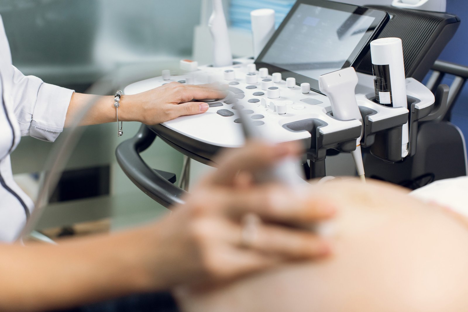
- For a tummy scan, a clear gel is placed on the skin and a small probe glides over your lower abdomen. Mild pressure helps obtain clear images.
- For a transvaginal scan, consent is confirmed, and a slim probe with a protective cover is placed gently in the vagina for closer views. A chaperone is available. You can ask to stop at any time.
- In pregnancy, we may measure the baby, check the anatomy, fluid, placenta, blood flow with Doppler, and baby position. In gynecology, we may measure the womb and lining, look for fibroids or polyps, examine the ovaries and tubes, and check for free fluid or signs of infection.
- Some results are available immediately. At times, images are reviewed by a second specialist, and a written report is issued. If further tests are needed, we explain why and how they help.
- We review the findings with you in clear terms and agree on the next steps, reassurance, follow-up, treatment, or referral when required.
Emergency State?
In urgent situations, contact immediately for help.
Available 24 Hours
+123-234-1234
Get Your Free Health Consultation or Book Your Appointment Now
When to seek urgent care
Go to hospital or call emergency services now if you have any of the following, heavy vaginal bleeding, severe or persistent lower belly or pelvic pain, fever with pelvic pain or foul discharge, a positive pregnancy test with pain or shoulder tip pain, fainting or dizziness, chest pain or shortness of breath, waters that are green or brown, or baby movements that stop or noticeably slow after 28 weeks.
Action plan
Bring your questions, previous reports, and a list of medicines and allergies.
After the scan, keep copies of your report and images for your records.
If you are pregnant, note any concerns such as bleeding, pain, or reduced movements, and mention them on arrival.
Book your appointment with the doctor to review results, understand what they mean for you, and plan follow-up or treatment if needed.
Wear two-piece clothing for easy access to the lower abdomen and consider bringing a sanitary pad in case of gel or spotting.
Follow any preparation advice; for some pelvic scans, a comfortably full bladder helps, for transvaginal scans, an empty bladder is usually best.
FAQ - Ultrasound in Obstetrics and Gynecology
Frequently Asked Questions
These FAQs offer general information for patients. They do not replace medical advice. For urgent concerns, contact your local emergency number or visit the nearest emergency department.
Yes. It uses sound waves, not radiation. It has been used in medical care for many years and is considered safe when medically indicated.
Transabdominal scans use a probe on the tummy, often with a partly full bladder. Transvaginal scans use a slim probe in the vagina, usually with an empty bladder, and give closer views. Consent is always requested.
It should not be painful. You may feel mild pressure. Your privacy is protected, a chaperone is available, and you can stop at any time.
Common scans include an early dating and viability scan, a nuchal translucency scan when offered, a detailed anatomy scan around the middle of pregnancy, and growth and well-being scans when needed.
Doppler measures blood flow, for example, in the umbilical cord or uterine arteries. It helps assess placental function and the baby well well-being.
No test can detect every condition. A normal ultrasound reduces risk and is very reassuring, yet some problems may not be visible, especially very small or late-developing findings.
Reasons include twins, growth concerns, high blood pressure, diabetes, reduced movements, bleeding, fluid changes, or placenta position.
Ultrasound helps confirm the location of the pregnancy and heart activity when the timing allows. If ectopic pregnancy is a concern, we act quickly and safely.
Yes. Ultrasound can locate IUD strings and device position, which helps guide care if there are symptoms or concerns.
As often as medically needed. We balance information and reassurance with the least number of visits required for safe care.
Heavy or irregular periods, pelvic pain, fibroids, polyps, ovarian cysts, suspected endometriosis, pelvic infection, and fertility assessment, including antral follicle count.
Instructions vary. Some pelvic scans work best with a comfortably full bladder. Transvaginal scans are usually done with an empty bladder. You will be advised what to do.
Yes, where space and privacy allow. For some parts of the scan, your companion may be asked to wait nearby.
Often yes. Ask the team. A formal report is sent to your clinician, and you can request a copy for your records.
Baby position, bowel gas, or body habitus can affect views. We may try different positions, schedule a repeat scan, or suggest complementary tests.
These can be helpful in selected clinical situations. Medical scans focus on information that guides care rather than entertainment images.
Yes. It can guide safe placement or removal of some devices, early pregnancy care, and drainage of certain cysts when appropriate.
Ultrasound gel is water-based and usually does not stain. Reactions are rare. Tell us if you have sensitive skin or allergies.
We explain the findings, answer your questions, and plan next steps. This may include repeat scanning, blood tests, referral, or treatment.